
The most potent cytotoxic payloads are often hydrophobic. Conjugated to a hydrophilic antibody, that hydrophobicity propagates into aggregation, accelerated systemic clearance, premature payload release and a narrower therapeutic window.
Our review organises hydrophilic linker design by position — attachment site, central spacer, release site — because the same polar motif behaves differently depending on where it sits in the architecture.
Challenges and Approaches to the Design of Hydrophilic Linkers for Antibody-Drug Conjugates
Potency Comes With a Hydrophobicity Problem.
Payload hydrophobicity is not a formulation detail — it shapes conjugate behaviour from manufacture through circulation. Hydrophilic linkers can offset these liabilities, particularly in higher-DAR designs, as one element of broader ADC optimisation rather than a fix on their own.
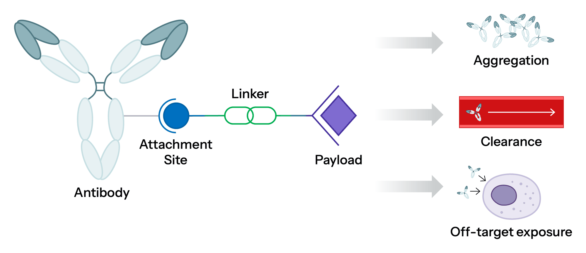
-
Aggregation
Reduced solubility and aggregation propensity as hydrophobic surface is exposed.
-
Clearance
Accelerated systemic clearance and nonspecific uptake.
-
Premature Release
Payload lost in circulation before the conjugate reaches the target.
-
Narrower Window
Off-target toxicity that constrains dose and DAR ambition.
Three Zones. One Architecture.
The review classifies hydrophilic linker strategies by the location of the solubilising element. This spatial view complements classification by motif type — PEG, zwitterions, carbohydrates, peptides — rather than replacing it.
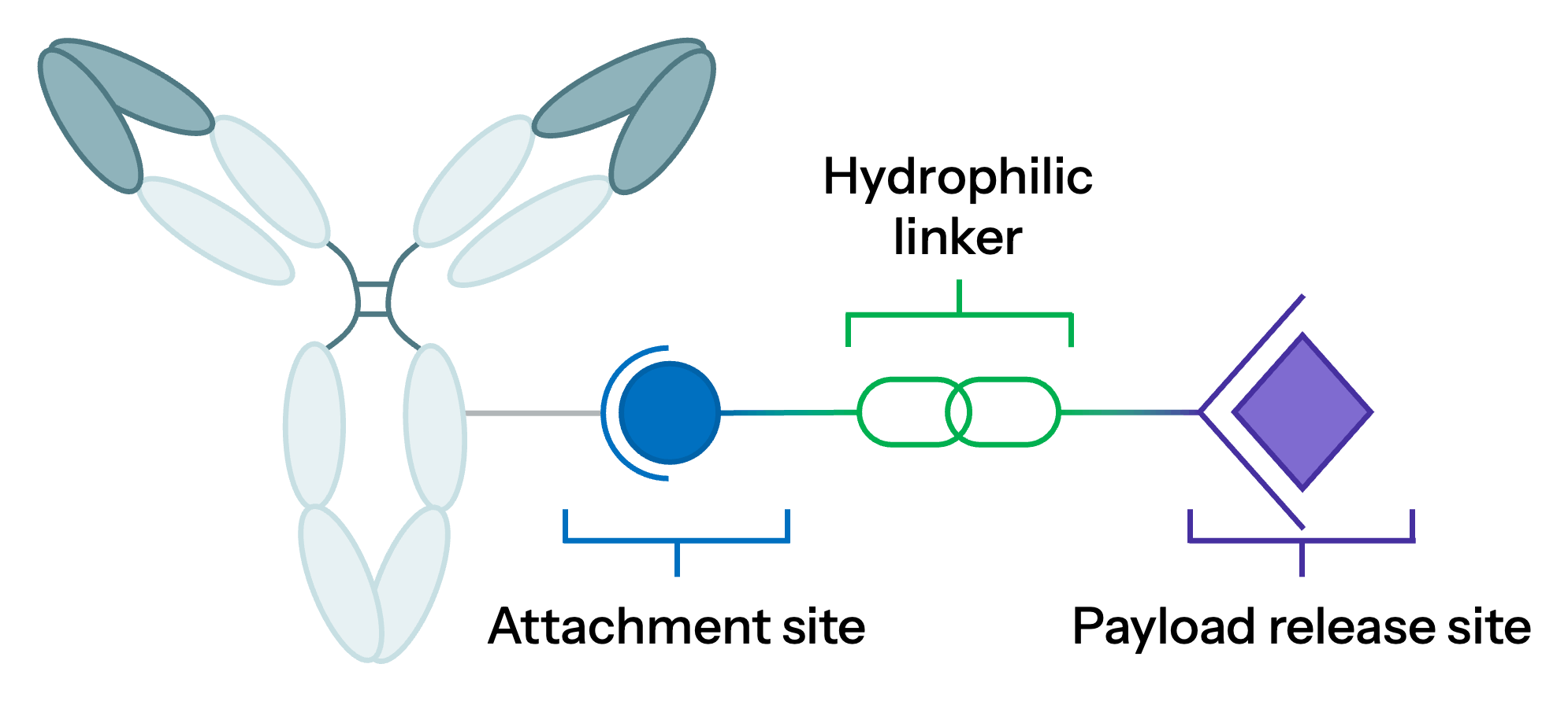
-
Zone 1 · Antibody Attachment Site
Improve local polarity and conjugation stability at the junction where the linker meets the antibody.
-
Zone 2 · Central Linker Spacer
Mask hydrophobic payload domains and improve overall developability across the conjugate.
-
Zone 3 · Payload-Release Site
Protect the linker–payload module in circulation and help tune intracellular release.
Key principle — the same hydrophilic motif can have different physicochemical and biological effects depending on its position and topology.
Build Stability and Polarity at the Conjugation Junction
Polar features near the attachment site reduce local hydrophobic clustering and improve conjugate stability.
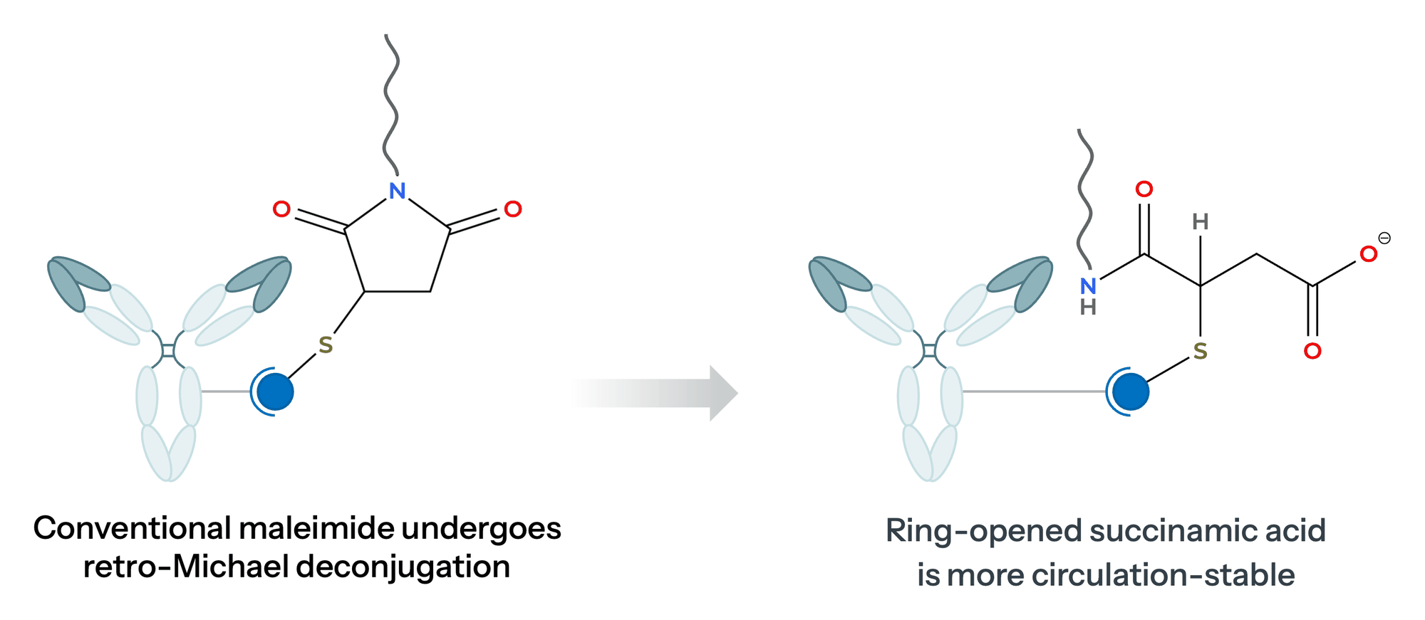
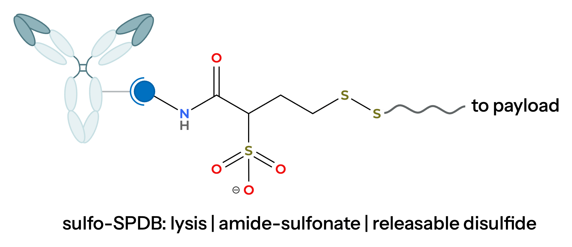
-
Self-stabilizing maleimides
Proximal basic amines or electron-withdrawing substituents promote hydrolysis to a stable, ring-opened succinamic-acid linkage — reducing retro-Michael deconjugation in circulation.
-
Charge modulation
Sulfonated linkers such as sulfo-SPDB raise local polarity and have been shown to reduce aggregation propensity and nonspecific clearance while retaining disulfide-mediated release.
Use the Spacer as a Hydrophilicity Reservoir
Chain length, position and three-dimensional presentation all matter. Performance depends on payload, DAR, antibody, conjugation site and the complete linker architecture.
-
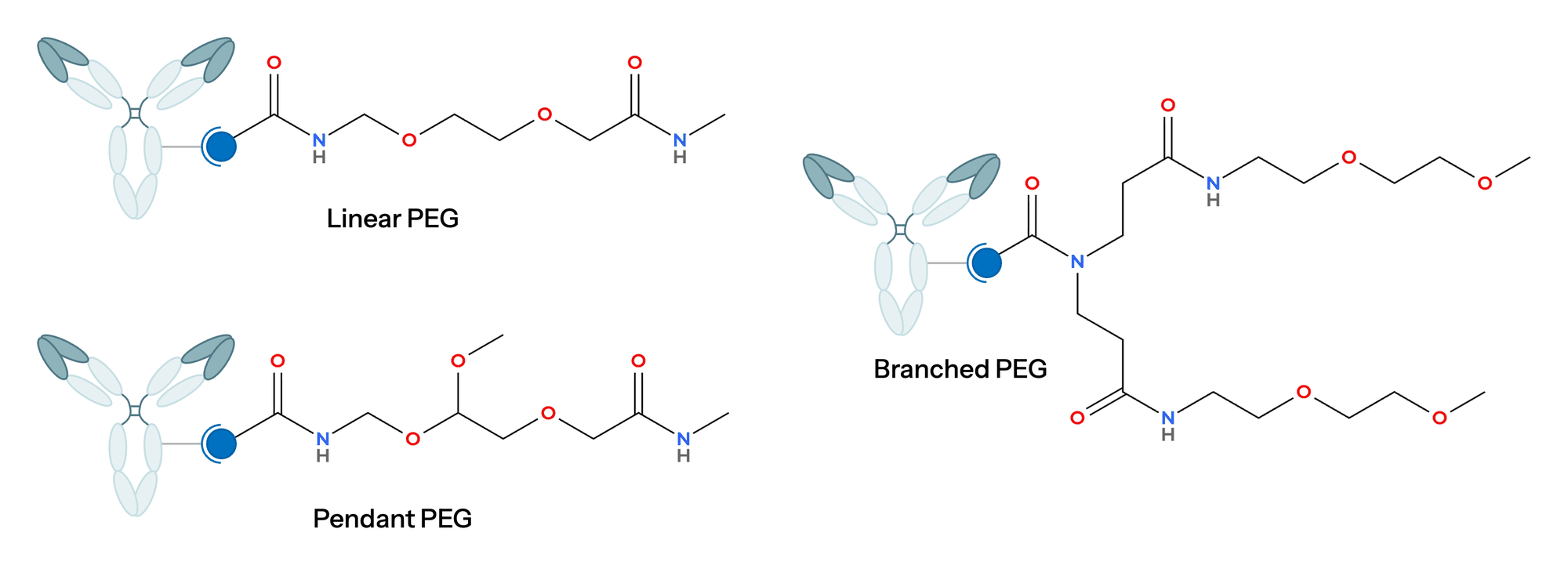
PEG architectures
PEG-containing linkers appear in clinical ADCs including Trodelvy and Zynlonta. In preclinical studies, pendant and branched-pendant designs shielded payloads more effectively than linear equivalents, particularly at high DAR.
-
Beyond PEG
Hydrophilic macrocycles — crown ether and cyclodextrin motifs — improved in vivo performance in reported studies. Acidic peptides such as Glu-Val-Cit and Asp-Val-Cit improved mouse plasma stability while preserving cathepsin-mediated cleavage.
Engineer Hydrophilicity at the Point of Release
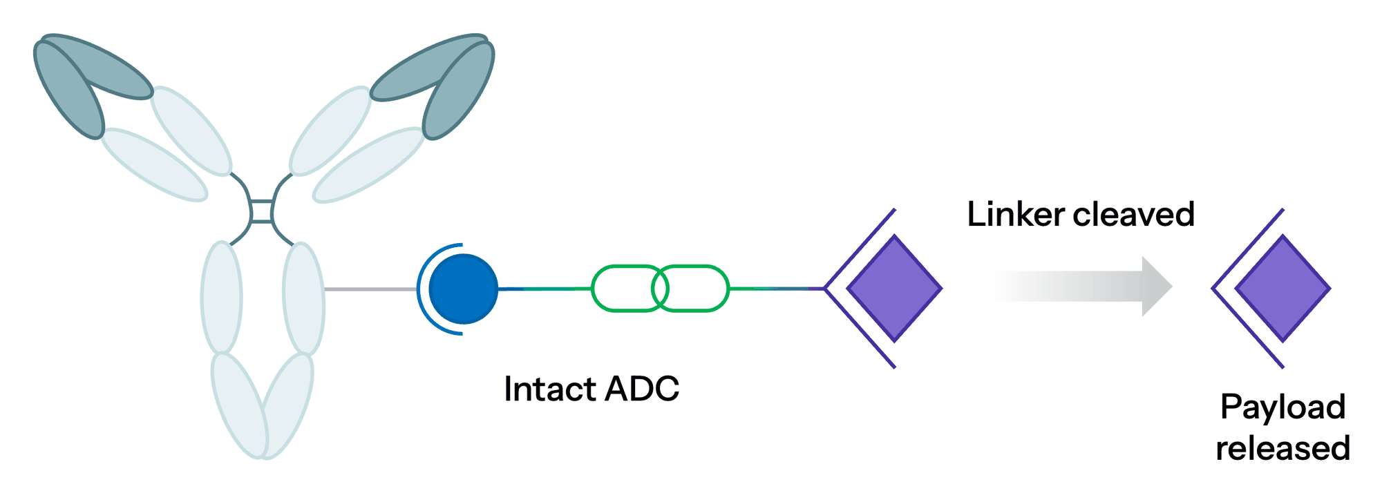
Polar groups near the cleavage trigger and self-immolative spacer mask the linker–payload module in circulation while supporting controlled intracellular release.
-
β-Glucuronide linkers
A dual-function design — β-glucuronide enhances hydrophilicity and acts as a substrate for β-glucuronidase after internalisation, releasing payload through a self-immolative cascade.
-
Quaternary ammonium linkers
Converting tertiary amine-containing payloads into permanently charged quaternary ammonium motifs can improve conjugate stability and hydrophilicity while preserving antibody-mediated delivery.
There Is No One-Size-Fits-All Hydrophilic Linker.
Hydrophilic linkers are no longer solubility modifiers. They are multifunctional design elements, optimised alongside the antibody, payload, conjugation chemistry, target DAR, release mechanism, target biology and manufacturability requirements.
Hydrophilic Linkers: Common Questions
What is a hydrophilic linker in an antibody-drug conjugate?
A hydrophilic linker is the chemical bridge between the antibody and the payload, built with polar or charged motifs such as PEG, zwitterions, carbohydrates or charged peptides. It offsets the hydrophobicity of potent cytotoxic payloads, helping limit aggregation, accelerated clearance and premature payload release, particularly in higher-DAR designs.
Why are hydrophobic payloads a problem for ADCs?
The most potent cytotoxic payloads are often hydrophobic. Conjugated to a hydrophilic antibody, that hydrophobicity can reduce solubility and promote aggregation, accelerate systemic clearance and nonspecific uptake, and raise the risk of premature payload release and off-target toxicity, which narrows the therapeutic window.
Where can hydrophilicity be placed in an ADC linker?
The review groups strategies into three zones. At the antibody attachment site, polar features improve local polarity and conjugation stability. In the central spacer, they mask hydrophobic payload domains. At the payload-release site, they protect the linker–payload module in circulation and help tune intracellular release.
Are PEG linkers always the best choice for ADCs?
Not universally. PEG-containing linkers appear in clinical ADCs including Trodelvy and Zynlonta, and pendant or branched PEG designs shielded payloads better than linear equivalents in preclinical studies, especially at high DAR. Performance still depends on payload, DAR, antibody, conjugation site and the complete linker architecture.
How does a β-glucuronide linker release its payload?
β-Glucuronide does two jobs. It adds hydrophilicity that helps mask the linker–payload module in circulation, and it acts as a substrate for β-glucuronidase once the ADC is internalised. Enzymatic cleavage then triggers a self-immolative cascade that releases the payload inside the cell.
Read the Review. Then Bring Us Your Molecule.
Our bioconjugation and ADC teams run linker design, conjugation chemistry, DAR characterisation and developability under one program — with the scientists who authored this work.
Ma X, Li B, Ma Y, Lau JYN. Challenges and Approaches to the Design of Hydrophilic Linkers for Antibody-Drug Conjugates. Journal of Controlled Release, 2026. PMID: 42668070.
