
Immunofluorescence (IF) staining is used to detect and localize antigens (e.g., proteins) in cells or tissue sections by using of fluorescent dye-conjugated antibodies or non-antibody methods of fluorescent staining (eg. DAPI). Compared with immunohistochemistry (IHC), IF is more widely used to detect co-localization of multiple targets with different fluorescent dyes.
ChemPartner offers high-quality and efficient services for IF full assay development, protocol validation, single/double staining, image collection, and data analysis/interpretation.
- Equipment: IHC autostainer (Dako AS Link48), fluorescence microscope with 100x oil lens (Zeiss, AXIO Scope A1), etc.
- Imaging software: ZEN 2011 (Zeiss), Image Pro Plus, Image J, etc.
- Antibodies with established methods
Almost all bright-field IHC staining protocols can be converted to IF staining with fluorescent antibodies.
- Tissue types: FFPE and frozen samples of cells and tissue, TMA
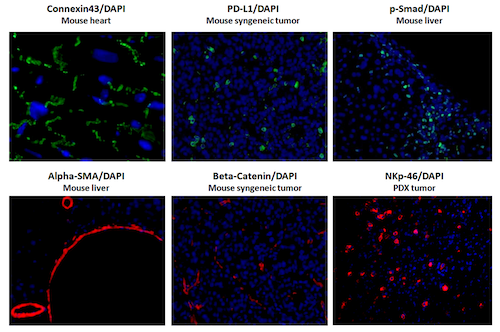
Representative Images of IF Staining
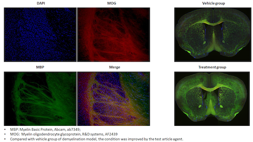
Representative Images of IF Double Staining
Deliverable:
- Raw data together with analyzed data in MS Excel format
- Images in JPEG format
- Project update and summary in MS PowerPoint format
- Final report in MS Word format (including purpose, methods, results, and data analysis)
Sample Submission: Test samples could be submitted
- FFPE blocks of tissues or cells
- Oct-embedded samples
- Tissues and cells fixed in formalin
- Fresh frozen samples
