
Immunofluorescence (IF) staining is used to detect and localize antigens (e.g., proteins) in cells or tissue sections by using of fluorescent dye-conjugated antibodies or non-antibody methods of fluorescent staining (eg. DAPI). Compared with immunohistochemistry (IHC), IF is more widely used to detect co-localization of multiple targets with different fluorescent dyes.
ChemPartner offers high-quality and efficient services for IF full assay development, protocol validation, single/double staining, image collection, and data analysis/interpretation.
- Equipment: IHC autostainer (Dako AS Link48), fluorescence microscope with 100x oil lens (Zeiss, AXIO Scope A1), etc.
- Imaging software: ZEN 2011 (Zeiss), Image Pro Plus, Image J, etc.
- Antibodies with established methods
Almost all bright-field IHC staining protocols can be converted to IF staining with fluorescent antibodies.
- Tissue types: FFPE and frozen samples of cells and tissue, TMA
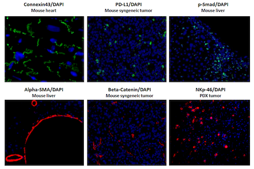
Representative Images of IF Staining
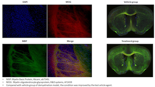
Representative Images of IF Double Staining
交付成果:
- 原始数据及分析后的数据(MS Excel格式)
- Images in JPEG format
- 以 MS PowerPoint 格式呈现的项目进展与总结
- MS Word格式的最终报告(包括研究目的、研究方法、研究结果和数据分析)
Sample Submission: Test samples could be submitted
- FFPE组织或细胞块
- Oct嵌入式样本
- 用福尔马林固定的组织和细胞
- 新鲜冷冻样本
