


Immunology and Inflammation Services for Translational Drug Discovery
Integrated in vitro, in vivo, and ex vivo immunology solutions to accelerate discovery and preclinical development across autoimmune, inflammatory, and immuno-oncology programs.
ChemPartner supports biopharma teams in evaluating immune function, characterizing candidate activity, and generating decision-ready data across development stages.
Our platform integrates primary immune cell assays, disease-relevant in vivo models, and translational ex vivo analysis to deliver both mechanistic insight and pharmacology-driven outcomes.
Integrated Immunology Capabilities
ChemPartner provides a connected immunology platform that bridges mechanism, efficacy, and translation through coordinated workflows across in vitro, in vivo, and ex vivo systems.
Our expertise spans both innate and adaptive immunity, including:
- Immune cell activation, proliferation, and differentiation
- Cytokine release and immune signaling pathways
- Receptor occupancy and ligand engagement
- Cell–cell interaction and co-culture systems
- Functional and phenotypic immune profiling
Core technologies include:
- Multi-color flow cytometry (FACS)
- ELISA, Luminex, ELISPOT
- QuantiGene transcriptomics
- AlphaLISA signaling assays
In Vitro Immunology Assays
Mechanistic and functional immune profiling across primary cells and complex co-culture systems.
Our in vitro platform enables highly customizable evaluation of immune responses across multiple cell types, pathways, and therapeutic modalities. Using whole blood, PBMCs, and primary immune cells, we generate robust functional and mechanistic data aligned to your target biology.
Immune Cell Function and Differentiation
We support detailed functional characterization across key immune populations:
- T cells: activation, proliferation, cytotoxicity, and killing assays
- Th subsets: Th1, Th2, Th17, and Treg differentiation
- Treg biology: suppressive function assays (human and mouse)
- B cells: activation and differentiation into plasmablasts/plasma cells
- NK cells: cytotoxicity and tumor cell killing (e.g., K562 models)
- Macrophages: M1/M2 polarization and phagocytosis
- Basophils: activation assays in human blood
Co-Culture and Functional Systems
Biologically relevant systems to evaluate immune engagement and mechanism:
- Tumor/T cell co-culture systems
- Macrophage/T cell co-culture
- Mixed lymphocyte reaction (MLR) assays
- CMV antigen recall assays
- OT-I T cell and OVA tumor co-culture models
- T cell engager (TCE) functional assays
Immune Signaling and Inflammation Pathways
Targeted assays for pathway interrogation and drug mechanism:
- JAK/STAT signaling assays (including phospho-STAT detection)
- NLRP3 inflammasome activation assays
- CD73–A2aR signaling pathway assays
- STING antagonist assays (human and mouse)
- TLR7/8-induced cytokine release assays
- LPS-induced cytokine release models (whole blood, PBMC, THP-1)
- Receptor occupancy and ligand-binding assays
Effector Function and Modality-Specific Assays
Support for complex therapeutic formats:
- ADCC, ADCP, CDC, TDCC
- Cytokine-targeting efficacy studies
- T cell engager (TCE) comprehensive studies
- Bi-specific antibody-mediated killing assays
Readouts and Analytical Outputs
Multi-dimensional data generation for mechanistic insight:
- Cytokine profiling (ELISA, Luminex, ELISPOT)
- Transcriptomic analysis (QuantiGene)
- Signaling pathway analysis (AlphaLISA)
- Multi-color flow cytometry (FACS)
- Phosphorylation-based signaling (pSTAT)
Case Study:
JAK Signaling Assay in Human Blood – Mechanistic pathway analysis using human blood and orthogonal readouts.
Representative human blood-based JAK signaling workflow showing cytokine-specific pathway interrogation with FACS and AlphaLISA readouts across defined immune-cell populations.
JAK signaling screening with validated PBMCS/blood pathways:
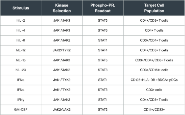
JAK signaling assay in human blood-FACS and ALPHALISA analysis:
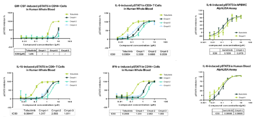

In Vivo Immunology and Inflammation Models
Disease-relevant models to evaluate efficacy, pharmacodynamics, and mechanism of action.
Our in vivo platform supports comprehensive pharmacological evaluation across a wide range of immune-mediated diseases. We offer:
- Conventional induced disease models
- Humanized and gene knock-in systems (e.g., hIL-4/IL-4R, hIL-17A/F)
- Over 40 customized pharmacodynamic (PD) models
- AAALAC-accredited research infrastructure
These models enable precise assessment of therapeutic efficacy, immune modulation, and mechanism of action.
Disease Areas and Representative Models
Respiratory Inflammation
- OVA- and HDM-induced asthma models
- Bleomycin-induced lung fibrosis
- LPS-induced acute lung injury
Rheumatoid Arthritis
- Collagen-induced arthritis (CIA)
- Adjuvant-induced arthritis (AIA)
Multiple Sclerosis
- EAE models (MOG, MBP, PLP systems)
Inflammatory Bowel Disease
- DSS-induced colitis (acute and chronic)
- TNBS-induced colitis
- T cell transfer models
- Humanized cytokine KI IBD models
Skin and Hypersensitivity
- Psoriasis models (IMQ, IL-23)
- Atopic dermatitis models (MC903, FITC, OXA/DNCB)
- Delayed-type hypersensitivity (DTH)
- Passive cutaneous anaphylaxis
- Arthus reaction models
Systemic Inflammation
- LPS-induced cytokine release
- Inflammasome activation (LPS + ATP)
- Con A-induced systemic inflammation
- Air pouch and CLP models
- SLE models
Case study
Type-II Collagen Induced Arthritis (CIA) in Rats – A well‑established in vivo model of rheumatoid‑like arthritis with clinical, imaging, and biomarker readouts.
Type-II collagen-induced arthritis model evaluated by HE staining, Safranin O staining, and x-ray across control, model, and compound groups, illustrating multi-endpoint assessment of inflammatory arthritis.
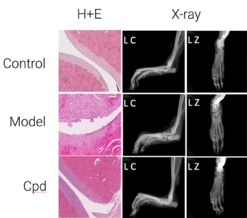
Type‑II collagen‑induced arthritis (CIA) in rats. Clinical scores, joint swelling, histology, and X‑ray images compare control, CIA model, and compound‑treated groups.
Request In Vivo Model Guidance
Ex Vivo Analysis Platforms
Translational analytics to connect in vivo results with mechanistic and biomarker insight.
Our ex vivo platform bridges biology and decision-making through advanced immunological analysis across diverse models, matrices, and modalities.
Core Capabilities
Immunophenotyping and Functional Biomarkers
- Immune cell profiling
- Functional biomarker assessment
- Tumor-infiltrating lymphocyte (TIL) analysis
- Multiplex cytokine analysis
PD/PK and Mechanistic Analysis
- Receptor occupancy
- Phosphorylation signaling (e.g., pSTAT)
- PK/PD correlation
Model Systems
- Syngeneic models
- Humanized (hPBMC, hCD34+) models
- Human gene knock-in models
- Wild-type animal models
Sample Types
- Whole blood
- PBMCs
- Lymph nodes, spleen, thymus
- BALF and bone marrow
Modalities and Species
- Modalities: antibodies, small molecules, XDCs, TCEs
- Species: human, NHP, mouse, rat, canine
Analytical Technologies
- FACS
- Luminex
- MSD
Customization
Custom panel design is available to align assays with:
- Target biology
- Mechanism of action
- Translational biomarker strategy
Case Study
TIL Analysis and Multi-Color Flow Cytometry – Ex vivo immune profiling to support translational biomarker and mechanism studies
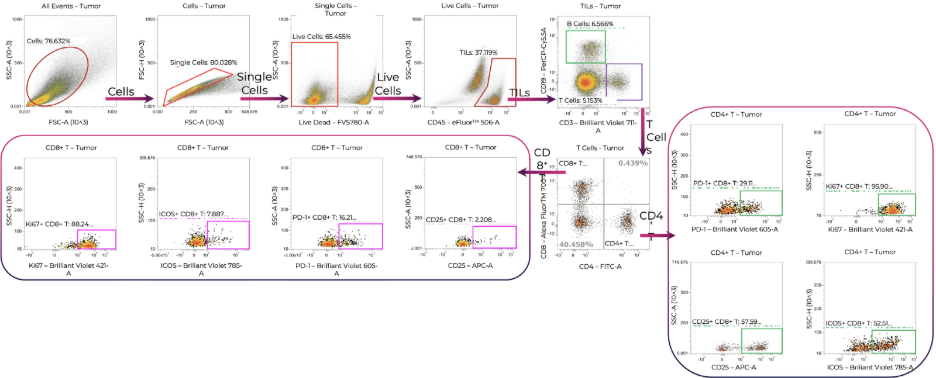
Legend: Representative ex vivo immune profiling workflow for checkpoint and activation marker analysis using multi-color flow cytometry.
Why ChemPartner
ChemPartner combines deep immunology expertise, disease-relevant pharmacology, and translational analytics within a unified platform.
- Integrated in vitro, in vivo, and ex vivo workflows
- Expertise across innate and adaptive immunity
- Broad autoimmune and inflammation model coverage
- Multi-species and multi-modality support
- Flexible PD and biomarker strategies
Advance Your Immunology Program
Whether you’re exploring early immune mechanisms or validating efficacy in complex disease models, ChemPartner helps design the right experimental strategy for your program.